


INJURIES OF THE ACCESSORY LIGAMENT OF THE DEEP DIGITAL FLEXOR TENDON IN HORSES: LOW FREQUENCY ULTRASOUND TREATMENT COMPARED TO A CONTROL CASE
DOTT.SSA Martina Palla, Equine Practitioner, Roma, Italia; Raffaella Maggi, DVM, Spec. Med. e Chir. Cavallo, Cert. AVP, MRCVS, Francesco Putti, DVM, Spec. Med. e Chir. Cavallo.
AIM OF THE STUDY:
The aim of this retrospective study is to evaluate the effects of Low Frequency Ultrasound (LFUS) therapy compared to a control horse, managed only with anti-inflammatory drugs, in the treatment of acute injuries of the proximal third of the accessory ligament of the deep digital flexor tendon (ALDDFT, Distal check ligament) in show jumping horses.
MATERIALS & METHODS:
Ultrasonographic examinations (US) of horses with ALDDFT injuries were reviewed. Inclusion criteria were the presence of acute (less than 7 days since first observation), unilateral lameness and diagnosis of an acute lesion within the third proximal of ALDDFT. Horses with multiple limb lameness or patients with concurrent lesions of the metacarpal region were excluded.
Clinical assessment of the horses was performed by the same experienced veterinary surgeon. The US examinations were performed using a GE logiq-V2 device with a linear probe 7.5 MHz (5-12 MHz); transverse and longitudinal section images were acquired.
All horses received a course of anti-inflammatory drugs (NSAIDs and steroidal drugs) for 5-7 days after the US diagnosis.
The LFUS device used for this study was an EQ-Ultrasound (BAC technology horse), which operates at 38 kHz (±2 kHz).
Horses that have been subjected to the LFUS treatment received 1-weekly session interval, for 4 weeks. LFUS treatment was started one week after the diagnosis of ALDDFT injury. The adopted protocol included the use of the grey transducer (flat transducer with pulsed emission) for 5 minutes at 30% of full power. Subsequently the trigger concave transducer was adopted for 7 minutes at 70% and then again 5 minutes of treatment with the flat transducer at 30% of full power.
Ultrasonographic reassessment of the lesion was performed for all patients 15 days after the end of the LFUS treatment or 21 days after the diagnosis and then 21 days after the second US check.
An appropriate rehabilitation and recovery plan was built for the first 4 weeks after the diagnosis: exercise included hand-walking for 10 minutes once a day for the first week increasing to 30 minutes of hand-walking at the end of the fourth week. After the 4th week the protocol was adapted for each case depending on the US examination. If the ALDDFT lesion was US improved, then an interval training protocol was adopted: from weeks 4 to 8 after the diagnosis, a gradual and increasing exercise plan including walking and trotting up to 20 minutes for each session. Canter exercise was gradually reintroduced for each patient.
RESULTS:
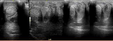
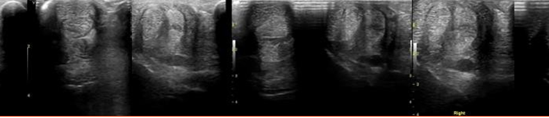
Three show jumping horses were included, all geldings with a median age of 18 years old. All patients had a 1/5 lameness (AAEP grading scale) and a visual and palpable swelling at the proximal metacarpus. The first US examination revealed an enlargement of the diameter of the ALDDFT and a hypoechoic area indicating oedema, haemorrhage and fibres damage within the ligament. Two patients received LFUS treatments and one of them was also treated with intralesional Platelet-Rich Plasma (PRP) prior to starting LFUS. The third horse was only managed with NSAIDs therapy, rest, and controlled exercise.
The first US reassessment showed a moderate improving in transverse section (mixed echogenicity: 50% echogenic and 50% anechoic) in the horses treated with LFUS, while in the other horse the lesion remained mostly anechoic. While in longitudinal images 50%-75% of fibres were aligned in LFUS treated patients and 25%-50% in the non-treated horse.
At the last US examination, the horses treated with LFUS showed in transverse section a lesion slightly hypoechoic, mostly echogenic, and the non-treated horse had still a mixed echogenicity lesion. In longitudinal section, more than 75% of the fibres were aligned in the first two horses, from 50% to 75% in the other one. The horses treated with LFUS were back at their previous level of exercise within 4 months, while the other one in 6 months.
CONCLUSIONS:
Desmitis of the ALDDFT is most frequently found in older show jumping horses (Boswell et al., 2010, Waguespack et al., 2011). The main limitation of this study is the small number of patients included although this is the first study about the use of LFUS in case of acute ALDDFT injuries. Carrozzo et al. (2019) described the use of LFUS for suspensory ligament injuries in horses.
Literature reports the use of NSAIDs and intralesional corticosteroids, eventually associated with shock wave therapy (Boswell et al.,2010), for ALDDFT injuries. Our results may represent a valid alternative approach with a decreasing of rehabilitation time compared to the horse treated with anti- inflammatory drugs only. Furthermore, LFUS are well tolerated by all patients, without the necessity of any sedation protocol.
REFERENCES:
• Boswell RP, Mitchell RD, Ober TR, Benoit PH, Miller CHB, and Dyson SJ. (2010) Lameness in the show hunter and in the show jumper. In P. Rudolph & L. Harms (Eds.), Diagnosis and Management of Lameness in the Horse (2nd ed., pp. 1097-1112).
• Carrozzo, U., Toniato, M., & Harrison, A. (2019). Assessment of Non-invasive Low-Frequency Ultrasound as a Means of Treating Injuries to Suspensory Ligaments in Horses: A Research Paper. J. Equine Vet. Sci, 80, 80–89.
• Waguespack RW, Burba DJ, Hubert JD, Vidal MA, Lomax LG, Chirgwin SR, Lopez MJ (2011) Effects of Extracorporeal Shock Wave Therapy on Desmitis of the Accessory Ligament of the Deep Digital Flexor Tendon in the Horse. Vet Sur (40) pp. 450–456.